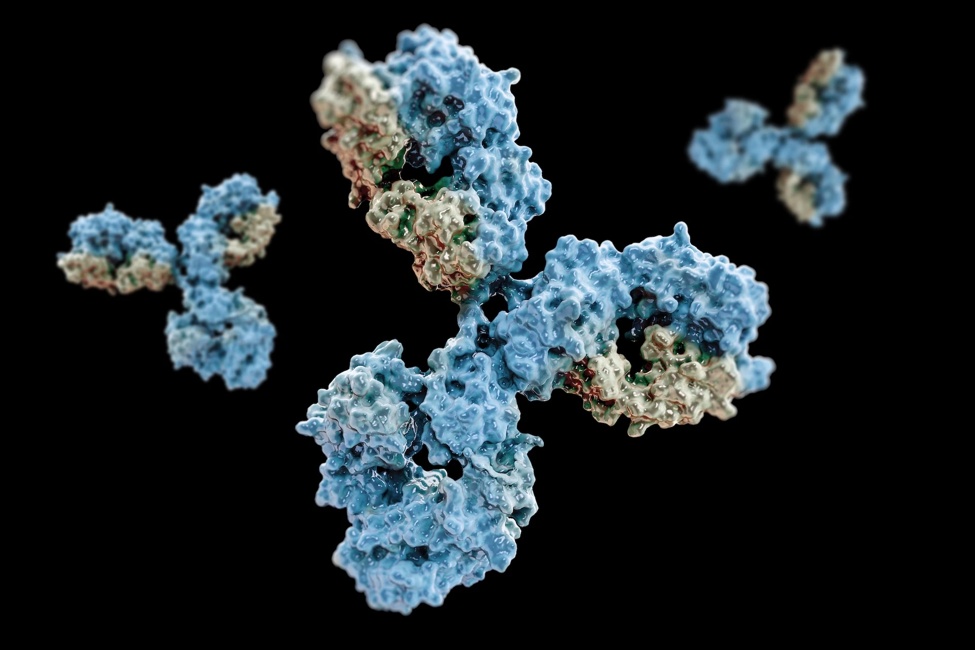
We probed β2-microglobulin (β2m) oligomers under Cu(II) using ESI–IM-MS and computational modeling. Monomers are uniform, but dimers/tetramers/hexamers show multiple conformations—tetramers are especially heterogeneous. This diversity correlates with Cu(II) release, a key step toward amyloid formation, indicating structural adaptability is functional (not a dead end); we also propose tetramer models that inform disease mechanisms and potential inhibitors.
Background
β2-microglobulin (β2m) aggregation drives dialysis-related amyloidosis, where misfolded proteins deposit in tissues and impair mobility. The earliest oligomers (dimers, tetramers, etc.) likely seed fibrils and toxicity but are hard to study because they’re fleeting and heterogeneous.
- Why early species are elusive: Short-lived, low-abundance, and structurally diverse → poorly captured by conventional structural methods.
- Role of metal ions: Cu(II) reshapes the β2m folding landscape and promotes oligomerization, complicating mechanism assignment.
- What we must resolve: The conformational diversity, dynamics, and metal-binding effects of pre-amyloid oligomers.
- Required toolkit: Techniques with both molecular specificity and shape/size resolution that can parse mixed populations in situ.
Challenge
Researchers needed to answer three key questions with molecular specificity and ensemble breadth:
- Cu²⁺-dependent architecture:
- How do β2m oligomers (dimers, tetramers, hexamers) assemble and organize in the presence of copper(II) ions—including metal-binding sites, oligomer stoichiometries, and shape/size distributions along the assembly pathway?
- Origins of heterogeneity:
- Why do higher-order species exhibit multiple, coexisting conformations while the monomer remains uniform? Are we seeing alternative interfaces, topologies, or mis-registered contacts, and how do solution conditions shift these conformational ensembles?
- Copper release → amyloidogenesis link:
- Does Cu²⁺ dissociation trigger conformational rearrangements that expose aggregation-prone surfaces, and on what timescales/kinetic steps does metal release couple to nucleation and fibril growth?
Traditional techniques (e.g., bulk spectroscopy, static structural methods) lack either the shape resolution, population sensitivity, or dynamic/kinetic visibility needed to map these transient, heterogeneous oligomer landscapes—necessitating approaches that capture structure, heterogeneity, and metal-coupled dynamics in tandem.
Approach: Molecular Simulation + Mass Spectrometry
Electrospray Ionization + Ion Mobility Mass Spectrometry (ESI–IM–MS)
- Native-like ESI preserved noncovalent assemblies, enabling detection of monomers, dimers, tetramers, and hexamers with their charge states.
- Ion mobility separations provided collision cross sections (CCSs) and conformer-resolved drift times, revealing coexisting shapes within each oligomer size.
- Comparative CCS analysis showed that structural heterogeneity peaks at the tetramer stage, marking a key branching point toward amyloid-competent architectures.
Molecular Simulation and Computational Modeling
- Built atomistic models for multiple tetrameric topologies and interfaces; evaluated their stabilities, packing, and exposure of aggregation-prone surfaces.
- Mapped Cu²⁺ coordination and release to specific conformational switches, showing how metal loss reweights the ensemble toward amyloidogenic geometries.
- Integrated CCS back-calculations and energetics to rationalize why diversity is functional—i.e., conformational adaptability supplies productive pathways to nucleation, rather than representing non-productive dead ends.
Key Findings
Uniform monomers, diverse oligomers
While β2m monomers adopt a single, consistent fold, dimers, tetramers, and hexamers populate multiple coexisting conformations, indicating early branching of assembly pathways.
Tetramers as the turning point
Conformational heterogeneity peaks at the tetramer, where distinct shapes and interfaces emerge. This stage is highly sensitive to Cu²⁺ binding/release, marking a key decision point toward amyloid-competent architectures.
Copper as a trigger
Cu²⁺ dissociation promotes rearrangements that expose aggregation-prone surfaces, thereby accelerating amyloidogenesis. Metal dynamics thus act as a molecular switch coupling oligomer structure to nucleation.
Simulation as a bridge
Computational modeling delivers atomistic tetramer models that explain the IM–MS trends (e.g., collision cross-section distributions) and rationalize why structural diversity is productive, not a dead end, in driving fibril formation.
Impact
For disease understanding
The study supplies a clear structural and mechanistic rationale for Cu²⁺-induced β2m amyloidogenesis: oligomer heterogeneity is not noise but a necessary conduit that steers ensembles toward amyloid-competent states. Identifying the tetramer as a heterogeneity peak reframes early aggregation as a productive, metal-coupled branching process.
For therapeutic design
Atomistic models of diverse tetramers reveal interfaces, pockets, and metal-linked switches that can be targeted to stall nucleation before fibril growth. This enables structure-guided inhibitor strategies (e.g., interface blockers, Cu²⁺-binding modulators, or conformational “trap” molecules) aimed at the earliest, most druggable nodes.
For molecular simulation promotion
By explaining ion-mobility and mass-spectrometric trends molecule-by-molecule, computational modeling elevates spectroscopy from observation to mechanistic inference. The result is a simulation–experiment feedback loop that turns heterogeneous signals into testable structural hypotheses, accelerating biomedical discovery beyond what either method can achieve alone.
Why It Matters
Amyloid diseases are propelled by fleeting, low-abundance oligomers that most experimental tools can’t isolate or resolve. Molecular simulation fills this gap by delivering atomistic, ensemble-aware models of these species—clarifying how metals like Cu²⁺ shift conformations, which interfaces seed nucleation, and what kinetic routes lead to fibrils. In turn, these mechanistic maps prioritize druggable targets, guide structure-based inhibitor design, and accelerate the path from observation to therapeutic strategy.
👉 Takeaway: By integrating molecular simulation with cutting-edge mass spectrometry, researchers revealed how copper-induced heterogeneity in β2m oligomers drives amyloid formation. This case underscores simulation’s power to turn hidden molecular events into actionable biomedical knowledge.
Reference
Structural heterogeneity in the pre-amyloid oligomers of β2-microglobulin.
Tyler M. Marcinko, Chungwen Liang, Sergey Savinov, Jianhen Chen, and Richard W. Vachet. J. Mol. Bio 432:396-409 (2019)


